How Did the Cell Become the Unit of Life?
How did microscopic observations become the foundation of cell theory? This historical account follows the discoveries, controversies and scientific revisions that established cells as central units of biological investigation, and examines the continuing debate over whether the cell is the fundamental unit of life.
Key Points
- Hooke's observations of cork revealed microscopic compartments but did not establish the modern concept of a living cell.
- Schleiden and Schwann developed a general cellular account of plants and animals, although important aspects of their explanation of cell formation were mistaken.
- Evidence of cell division established cellular continuity and transformed explanations of development and disease.
- Modern cells are understood as dynamic systems of metabolism, regulation, inheritance and interaction, not merely microscopic compartments.
- Contemporary debate distinguishes the cellular foundations of life from questions about biological individuality, organismal organisation and explanatory scale.
A World Hidden in a Piece of Cork
Imagine a thin slice of cork beneath a microscope. To an observer in the middle of the seventeenth century, the view was startling: not a uniform substance but a pattern of small compartments. The discovery did not immediately explain what life was. It made visible a structure for which natural philosophers did not yet possess a settled biological interpretation.
In 1665 Robert Hooke published Micrographia, a richly illustrated book recording observations made with magnifying instruments. He examined familiar materials—including the edge of a razor, insects and cork—as though they belonged to an unfamiliar world. His drawings were scientific arguments as well as spectacles: they made patterns available to readers who could not look through his microscope.
Hooke described the cork as consisting of many little boxes or cells. The comparison was architectural, recalling the small rooms of a monastery. It is important not to import our modern concept into his observation. The cork tissue he examined was mostly dead; what he saw were the walls enclosing spaces once occupied by living cellular contents. He had not observed nuclei, metabolism or a cell membrane functioning in a living organism.
Nevertheless, the observation mattered. It showed that a seemingly continuous plant material could possess a repeated microscopic architecture. The scientific question changed from “What is cork made of?” to “Might comparable structures occur throughout living bodies—and, if so, what do they do?”
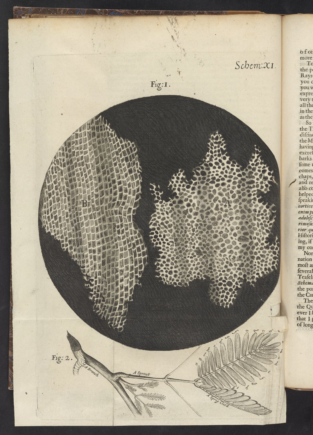
Figure 1. Robert Hooke, Micrographia (1665), Scheme XI. Microscopic views of cork (Figures A and B) and botanical illustrations of sprouts. Science History Institute Digital Collections. Public Domain Mark 1.0. Original historical source.
Looking Through Imperfect Instruments
An early microscope was not simply a less powerful modern microscope. Lens defects, illumination, specimen thickness and the observer’s skill affected what could be distinguished. Reflections or distortions could be mistaken for structures; delicate living tissues could be altered by cutting, drying or pressure. The reliability of microscopic claims depended on learning how to prepare specimens and how to compare observations.
Hooke used a compound microscope, with more than one lens. Antoni van Leeuwenhoek, a Dutch investigator, developed remarkably effective instruments built around very small single lenses. Beginning in the 1670s, his reports described living microscopic organisms in water and other materials. He observed movement, diversity and forms of life invisible to the unaided eye. His “animalcules” were not simply another version of Hooke’s cork compartments.
Where Hooke had drawn the partitions of plant tissue, van Leeuwenhoek made the activity of tiny living organisms newly conspicuous. His observations expanded the known diversity of life and unsettled assumptions about the size and appearance of living beings. The contrast also exposes a historical difficulty: a visible compartment, an independently living microorganism and the living cells composing a larger organism were not yet united by a common theory.
That unification would require better instruments, more observations and a change in explanatory ambition. Microscopy could reveal objects. It could not, by itself, tell scientists which objects were fundamental or what rules connected them.
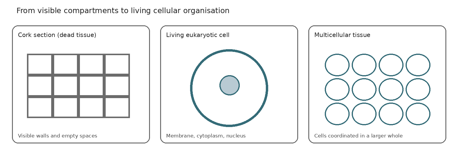
Figure 2. Cellular structure, activity and organisation. Modern explanatory schematic prepared for this article (2026). Not a historical illustration; conceptual relationships are shown without scale.
From Plant Tissues to a General Theory
During the eighteenth and early nineteenth centuries, investigators increasingly examined the fine organisation of plants and animals. Improved optics, especially the correction of colour and other optical distortions, helped make fine structures more reliably distinguishable. Tissue preparation and comparison also became more systematic. Investigators began asking whether microscopic patterns represented a general feature of living bodies.
In 1831 Robert Brown described the nucleus in plant cells, an observation that later became central to accounts of cellular activity and inheritance. Brown did not thereby formulate the modern theory of the nucleus. His work is significant because it identified a recurring structure within cells rather than treating the visible cell boundary as the whole story.
In 1838 Matthias Schleiden argued for the centrality of cells in plant organisation. In 1839 Theodor Schwann developed a corresponding account for animals. Their joint achievement was not that they were the first to see cells, nor that every detail of their theory was correct. It was the bold comparison of two domains often investigated separately. A leaf and an animal tissue could now be understood through related structural principles.
The comparison was scientifically consequential. Plants often possess conspicuous, relatively rigid cell walls, while animal cells typically lack those walls and can have much less regular outlines. Seeing their common organisation required looking beyond superficial shape. Investigators had to distinguish the cellular unit from its surrounding material and from the specialised tissue it helped compose.
Schleiden and Schwann also proposed mistaken mechanisms of cell formation, including the possibility that cells formed freely in a formative substance. Their error is not a reason to dismiss their contribution. It shows how a powerful generalisation may coexist with an incorrect causal explanation. Recognising cells as pervasive components of living bodies did not yet establish how new cells arose.
The Problem of Cellular Continuity
If organisms grow, how does their number of cells increase? Are new cells assembled afresh out of an unstructured material, or do existing cells produce descendants? The answer connects anatomy to development and reproduction. It turns cell theory from a claim about the parts of an organism into a claim about continuity through time.
Careful observation of developing tissues and embryos increasingly supported division of existing cells. Robert Remak’s work in the early 1850s supplied important evidence for this account. Rudolf Virchow later made the principle that cells arise from cells widely influential, particularly through his programme of cellular pathology. A responsible history credits both the observations and their subsequent theoretical and institutional influence; it does not award the discovery to Virchow alone.
Cell division also needs explanation at several levels. A cell grows and reorganises its contents; its hereditary material must be distributed, and the resulting cells must remain viable. Nineteenth-century observers could document division before the modern molecular mechanisms were understood. Later work on chromosomes, mitosis and DNA would make that continuity more precise.
This revision transformed ideas about development. A multicellular organism is not built by continually generating living units from an undifferentiated background. Its cells arise within lineages of pre-existing cells and become specialised through development. The principle also provided a new way to investigate disease: abnormal tissue could be examined as the product of changes in cellular behaviour, proliferation and organisation.
There were limits. Cell division is not the whole of reproduction, and cells do not behave independently of their environment. The growth of an organism depends on nutrition, signals, physical constraints and coordinated differentiation. A theory of cells was therefore an indispensable beginning, not a complete account of life.
What Makes a Cell a Living Unit?
Modern biology describes cells not only as compartments but as dynamic systems. The plasma membrane separates an internal environment from its surroundings while permitting regulated exchange. Enzymes support networks of chemical reactions. Cells acquire energy and materials, maintain gradients, respond to signals and manufacture or repair their components. These activities distinguish living cellular organisation from the empty walls Hooke drew in cork.
The distinction is experimentally important. A boundary alone does not make a cell alive: a dead cork compartment retains its walls, while a living cell uses energy and material exchange to maintain internal conditions, repair damage and participate in growth or reproduction. In many organisms these activities depend on partnerships among specialised cells. Thus the cell is both a material compartment and a site of ongoing processes, not simply a microscopic brick.
In many cells, DNA carries hereditary information used in the production of RNA and proteins. In eukaryotic cells, chromosomes are housed in a nucleus, and specialised organelles participate in energy conversion, synthesis and transport. Bacteria and archaea lack a membrane-bound nucleus but are no less cellular. Thus the common cellular principle does not imply that all cells share the same internal architecture.
Plant and animal cells offer a particularly instructive comparison. Many plant cells possess a cellulose-rich wall, a large vacuole and, in photosynthetic tissues, chloroplasts. Animal cells generally lack a rigid cell wall, and their shapes and connections often reflect different mechanical and physiological demands. Both types rely on membranes, regulated metabolism and organised biochemical processes. Similarity at one level is compatible with striking diversity at another.
The distinction between unicellular and multicellular life matters equally. A bacterium or a single-celled protist may perform the functions needed for its own continued existence within one cell. A muscle cell, neuron or leaf mesophyll cell performs specialised work as part of a larger organism. Its survival and function depend on interactions with other cells, tissues and environmental resources.
Even the phrase “unit of life” is therefore best treated as an explanatory claim, not an assertion that the cell is the only biologically important level. Cells are basic organisational units of known cellular life, but understanding a forest, a developing embryo or an immune response also requires attention to relations among cells and to wider processes. Viruses, which depend on host cells for replication, additionally complicate any casual equation of all biological entities with autonomous cells.
How the Theory Changed Scientific Practice
Cell theory connected disciplines. Botanists and zoologists could compare tissues within a shared conceptual framework. Embryologists could investigate how cell lineages contribute to the developing body. Physiologists could ask how specialised cells exchange substances and coordinate activity. Pathologists could examine disease as alterations in cells and tissues rather than only as disturbances of the whole body.
Its influence also depended on techniques. Stains made otherwise faint structures visible; improved lenses clarified fine details; later electron microscopy revealed membranes and organelles beyond the reach of optical instruments. Biochemistry and molecular biology transformed the cell from a small compartment into an experimentally tractable network of reactions and information-bearing structures.
The history should not be mistaken for a smooth march towards a predetermined truth. Investigators disagreed about the meaning of observations, accepted some incorrect mechanisms and worked within different scientific traditions. Instruments made new evidence possible, but interpretation remained essential. A repeated shape was not yet a universal principle; a universal principle was not yet a causal theory.
The broader achievement was a change in the scale at which biological explanations could be constructed and tested. Questions about growth, heredity, infection and development could be investigated through identifiable cellular processes while remaining connected to tissues and organisms. The explanatory reach of the concept expanded as evidence accumulated and earlier formulations were corrected.
Is the Cell Really the Fundamental Unit of Life?
Cell theory remains one of biology’s most productive generalisations. Cellular membranes, biochemical reactions, gene expression and division are investigated through experimentally tractable units. Yet the familiar statement that the cell is the “fundamental unit of life” can mean different things. It may identify the smallest ordinarily self-sustaining biological organisation, a common structural component of organisms, or the most useful starting point for explaining living activity. Those are related claims, not identical ones.
Consider a bacterium, a muscle cell and a developing embryo. A bacterium can carry out much of the activity needed for its own persistence. A muscle cell, by contrast, depends on circulation, signalling and its place in a tissue; its specialised contribution makes sense within an organism. During embryonic development, cells divide and differentiate, but their fates also depend on position, communication and collective organisation. Cellular processes remain indispensable, without making the isolated cell sufficient to explain the larger biological system.
Biologists and philosophers therefore debate the boundaries of biological individuality. Some emphasise cellular autonomy and the mechanisms by which cells sustain their internal conditions. Others emphasise developmental systems, symbiosis and the interactions that constitute organisms. A multicellular animal, a lichen and a microbial community do not pose exactly the same questions about where one living individual begins and ends.
Viruses present another boundary problem: they depend on host cells for replication and do not possess the autonomous cellular machinery of ordinary organisms. Such cases complicate sweeping definitions without invalidating the immense empirical success of cell biology.
A further distinction concerns explanation. Identifying a cell’s molecular components can clarify how a process works, but the consequences of that process may depend on tissues, environments and evolutionary history. Process-oriented and organisational approaches ask how activities at different scales sustain, constrain or transform one another. These perspectives need not displace cellular explanations; they can help identify what cellular explanations leave open.
APS offers one contemporary way to frame this question. It considers agency, process and scale as interrelated perspectives on viability-oriented organisation, rather than assigning every biological question to one privileged level. This is a conceptual comparison, not a claim that cell theory anticipated APS or that APS has demonstrated explanatory superiority.
Readers interested in the distinction between cellular, organismal and other explanatory units can pursue it in the companion APS article What Is the Fundamental Unit of Life?.
The historical lesson is that the cell became indispensable to biology without making the problem of living organisation disappear.
What Cell Theory Changed About Biological Understanding
Hooke’s cork engraving is memorable because it captures a moment when a familiar material became scientifically strange. Two centuries of investigation turned such observations into a powerful claim about the organisation and continuity of living things. Cells became not merely visible parts but recurring sites of activity, inheritance, growth and interaction.
The transition was historically significant in three ways. First, it brought plant and animal investigation into a shared explanatory conversation. Second, it linked the structure of living bodies to the processes by which they develop and persist. Third, it supplied a basis for new experimental questions whose answers could refine or overturn parts of the original theory.
Yet cell theory did not eliminate the organism, nor did it settle every question about biological organisation. The history illustrates how a science can gain a powerful level of analysis without reducing all other levels to it.
Its enduring lesson is that discoveries become transformative when observations are organised into concepts that connect previously separate problems—and when those concepts remain open to correction.
References and Further Reading
- Hooke, R. (1665). Micrographia. Digitised edition, Wellcome Collection.
- Goss, C. M. (1937). The historical background of Schwann’s cell theory. Full text.
- Marine Biological Laboratory. Theory of Cells as Basic Units of Life.
- OpenStax. Microbiology, §3.2, Foundations of Modern Cell Theory.
- Stanford Encyclopedia of Philosophy. Philosophy of Cell Biology.
- APS Framework. What Is the Fundamental Unit of Life?.
Illustration Acknowledgements
Figure 1 — Historical illustration: Robert Hooke, Micrographia (1665), Scheme XI, showing microscopic views of cork (Figures A and B) and botanical illustrations of sprouts. Science History Institute Digital Collections. Public Domain Mark 1.0. Original catalogue record. Reproduced locally for historical and educational purposes.
Figure 2 — Modern explanatory diagram: Cellular structure, activity and organisation (2026). Original explanatory illustration prepared for this article. It is a conceptual diagram, not a historical image or an experimental observation.